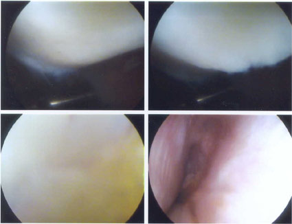
Top Left: A probe is tapping on bare bone (the top of the tibia, where
the tibia meets the knee) that should be covered with cartilage but isn't.
Top Right: Probe is not in the picture. You can see the hole where
the cartilage should be.
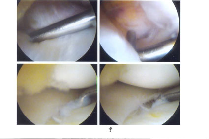
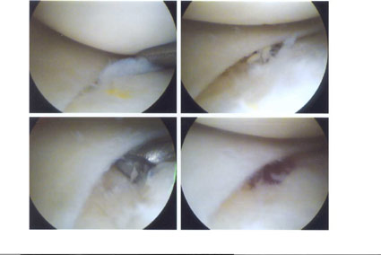
Bottom Left: A different instrument is used to actually do the microfracture.
This instrument is making tiny holes in the bone.
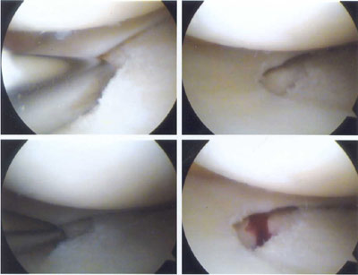
Bottom Right: The bone is bleeding after the holes were made. The hope
is that a scab will form and then the bone will send new cells to the area
and a new surface will be formed on top of the bone.